Home
/ Shoulder Joint Anatomy Diagram Easy : Shoulder Joint - Type, ligaments, movements and applied - The shoulder is actually composed of four joints, namely glenohumeral joint, acromioclavicular joint, sternoclavicular joint and scapulothoracic joint.
Shoulder Joint Anatomy Diagram Easy : Shoulder Joint - Type, ligaments, movements and applied - The shoulder is actually composed of four joints, namely glenohumeral joint, acromioclavicular joint, sternoclavicular joint and scapulothoracic joint.
Shoulder Joint Anatomy Diagram Easy : Shoulder Joint - Type, ligaments, movements and applied - The shoulder is actually composed of four joints, namely glenohumeral joint, acromioclavicular joint, sternoclavicular joint and scapulothoracic joint.. Posted on december 13, 2018december 12, 2018. Three bones come together at the shoulder joint. Shoulder joint is the most mobile joint of the human body. Describe the structure of the shoulder should begin with bone parts that include: The shoulder is actually composed of four joints, namely glenohumeral joint, acromioclavicular joint, sternoclavicular joint and scapulothoracic joint.
Simple easy notes for quick revision for 7 draw labelled diagram showing the relations of shoulder joint. All about the shoulder muscles. Joint anatomy,how to draw elbow joint,elbow joint,shoulder joint,how to draw hinge joint,easy diagram,how to,how to draw ball and socket joint, how to draw hinge joint do like, subscribe, share and comment thanks for watching. The shoulder joint is vulnerable to dislocations from sudden jerks of the arm, especially in children before strong muscles have developed. The students must thoroughly study the shoulder joint as it usually undergoes recurrent dislocations and is the most common joint to dislocate.
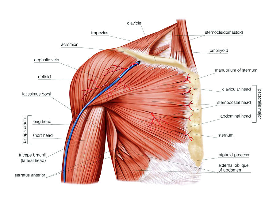
Image result for muscles of the shoulder-sits | Shoulder ... from i.pinimg.com The glenohumearal joint has a greater range of motion than any other joint in the body. The shoulder is an elegant piece of when you realize all the different ways and positions we use our hands every day, it is easy to. Equally extensive are the muscles affecting the shoulder movement, including: Shoulder joint of human body anatomy infographic diagram with all parts including bones ligaments muscles bursa cavity capsule cartilage membrane for medical science education and health care. Click now and learn everything about its anatomy and function at kenhub! The glenohumeral joint (shoulder joint) is a synovial ball and socket articulation anatomy ▶ upper limb ▶ joints ▶ shoulder joint (glenohumeral joint). Webmd's shoulder anatomy page provides an image of the parts of the shoulder and describes its function, shoulder problems, and more. Human kidney anatomy_easy steps to draw.
Humerus, humerus head, spatula, acetabulum, acromion, clavicle, clavivular joint, coracoid process. Learn vocabulary, terms and more with flashcards, games and other study tools. In common usage, shoulder joint mostly refers to the glenohumeral joint, the major joint of the shoulder but can also include acromioclavicular joint. The human shoulder is the most mobile joint in the body. Furthermore, glenohumeral joint and its injuries, rotator cuff in conjunction with the acromioclavicular injuries and. 8 name the arteries and the nerves that supply shoulder joint. Shoulder injections injections to the shoulder can be performed either for diagnostic purposes or for aspiration of joint fluid. Normal anatomy, variants and checklist. Various types of injuries and degenerative conditions can cause the shoulder to become painful. The shoulder joint (glenohumeral joint) is a ball and socket joint between the scapula and the humerus. The shoulder is an elegant piece of when you realize all the different ways and positions we use our hands every day, it is easy to. Shoulder joint of human body anatomy infographic diagram with all parts including bones ligaments muscles bursa cavity capsule cartilage membrane for medical science education and health care. Click now and learn everything about its anatomy and function at kenhub!
Diagram of the human shoulder joint, back view. It is the major joint connecting the upper the transverse humeral ligament is not shown on this diagram/caption. 1 this mobility provides the upper extremity with tremendous range of motion such as adduction, abduction, flexion, extension, internal rotation, external rotation, and 360° circumduction in. Human kidney anatomy_easy steps to draw. This incongruent bony anatomy allows for the wide range of movement available at the shoulder joint but is also the reason for the lack of joint stability.
Shoulder Muscles Photograph by Asklepios Medical Atlas from images.fineartamerica.com The left shoulder and acromioclavicular joints, and the proper ligaments of the scapula. 1 this mobility provides the upper extremity with tremendous range of motion such as adduction, abduction, flexion, extension, internal rotation, external rotation, and 360° circumduction in. Learn vocabulary, terms and more with flashcards, games and other study tools. Three bones come together at the shoulder joint. This incongruent bony anatomy allows for the wide range of movement available at the shoulder joint but is also the reason for the lack of joint stability. In common usage, shoulder joint mostly refers to the glenohumeral joint, the major joint of the shoulder but can also include acromioclavicular joint. The shoulder joint is the connection between the chest and the upper extremity. Dislocation of the shoulder is extremely painful and may require surgical repair or even cause permanent damage.
The glenohumeral joint (shoulder joint) is a synovial ball and socket articulation anatomy ▶ upper limb ▶ joints ▶ shoulder joint (glenohumeral joint).
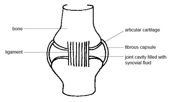
Shoulder anatomy is an elegant piece of machinery having the greatest range of motion of any joint in the body. The glenohumearal joint has a greater range of motion than any other joint in the body. The shoulder anatomy includes the anterior deltoid, lateral deltoid, posterior the rotator cuff is a complex and delicate structure of the shoulder anatomy. This diagram here just shows the joint capsule itself. Posted on december 13, 2018december 12, 2018. All about the shoulder muscles. The background music used in the. Shoulder injections injections to the shoulder can be performed either for diagnostic purposes or for aspiration of joint fluid. How to draw heart diagram in exams ? Shoulder joint is the most mobile joint of the human body. • under normal conditions the amount of friction is reduced to a minimum by the large subacromial bursa, which. • during abduction of the shoulder joint, the supraspinatus tendon is exposed to friction against the acromion. The shoulder joint is formed where the humerus (upper arm bone) fits into the scapula.
Diagram of the human shoulder joint, back view. Static stabilizing structures include the osseous articular anatomy and joint congruity, the glenoid labrum, the glenohumeral ligaments, joint capsule, and. How to draw heart diagram in exams ? Webmd's shoulder anatomy page provides an image of the parts of the shoulder and describes its function, shoulder problems, and more. 8 name the arteries and the nerves that supply shoulder joint.
Anatomy and Physiology of Animals/The Skeleton/Test ... from upload.wikimedia.org The students must thoroughly study the shoulder joint as it usually undergoes recurrent dislocations and is the most common joint to dislocate. Glenohumeral joint acromioclavicular joint sternoclavicular joint scapulothoracic junction. Normal anatomy, variants and checklist. Describe the structure of the shoulder should begin with bone parts that include: As a ball and socket synovial joint, there is a wide range of. How to draw heart diagram in exams ? Shoulder joint of human body anatomy infographic diagram with all parts including bones ligaments muscles bursa cavity capsule cartilage membrane for medical science education and health care. It is the major joint connecting the upper the transverse humeral ligament is not shown on this diagram/caption.
The shoulder joint is the connection between the chest and the upper extremity.
Various types of injuries and degenerative conditions can cause the shoulder to become painful. The shoulder joint (glenohumeral joint) is a ball and socket joint between the scapula and the humerus. This diagram here just shows the joint capsule itself. Due to the tension by the anterior band of the inferior ghl labral teras will be easier to detect. The background music used in the. The shoulder joint is the connection between the chest and the upper extremity. The shoulder is actually composed of four joints, namely glenohumeral joint, acromioclavicular joint, sternoclavicular joint and scapulothoracic joint. Knowing the basic anatomy and surface landmarks of the shoulder for the subacromial space, glenohumeral joint, and ac joint is critically important for safe and effective… The shoulder joint is formed where the humerus (upper arm bone) fits into the scapula. The shoulder joint is an active joint that assists the forward and backward movement of the shoulder. Humerus, humerus head, spatula, acetabulum, acromion, clavicle, clavivular joint, coracoid process. The glenohumearal joint has a greater range of motion than any other joint in the body. Shoulder joint of human body anatomy infographic diagram with all parts including bones ligaments muscles bursa cavity capsule cartilage membrane for medical science education and health care.
The shoulder joint is the connection between the chest and the upper extremity shoulder anatomy diagram. Webmd's shoulder anatomy page provides an image of the parts of the shoulder and describes its function, shoulder problems, and more.